07958 483 682
 p.resteghini@nhs.net
 For NHS appointments 0208 510 7871
Case Studies
Physiotherapy and Sports Injury Clinic based in E9, London
 Tarsal Tunnel: Demonstration of the Appearance of Nerve, Artery and Tendon
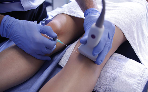
Hip joint: anterior approach intra-articular injection
Knee joint: injection of an effused suprapatellar pouch
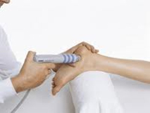
Forearm: Proximal intersection
Knee joint: Large loose body in the lateral suprapatellar pouch
Patient Presentation: Distance Runner
A 46 year old female distance runner. The patient had recently switched to a very low profile unsupportive running shoe during her build up to a Marathon. Within 2 weeks she had noted a severe pain at the medial aspect of the ankle and was unable to continue running.
Ultrasound findings & Management
Ultrasound of the medial ankle demonstrated a marked tenosynovitis with synovial thickening within the tibialis posterior tendon sheath around the posterior aspect of the medial malleolus. The tibialis tendon itself demonstrated no evidence of significant tendinopathy or tear. Treatment consisted of analysis of foot position and gait. A pronated foot type being noted. A more supportive running shoe was advised with a relatively rigid mid-sole. Orthotics were supplied for everyday footwear to off-load the tendon. In addition after consenting and under ultrasound guidance the tendon sheath of tibialis posterior was injected with 25mg hydrocortisone and 5mls 1% local anaesthetic. The patient was advised appropriate rehabilitation and on a slow return to running over the next 2 week. The patient reported that within 1 month they had increased their running to their previous levels.
Patient Presentation: Fighter's Thumb
A 38 year old male Mixed martial artist who twisted the right thumb while fighting 4 weeks previously. He had attended his local Emergency Department and was advised on rest and ice. XR taken at this time had demonstrated no bony injury.
After 1 month his pain persisted and his GP had referred him to our Sports Medicine Clinic. Examination demonstrated tenderness around the ulnar aspect of the right thumb at the level of the metacarpophalangeal joint with a sense of increased laxity.
Ultrasound Findings & Management:
Ultrasound of the Ulnar Collateral Ligament (UCL) of the thumb at the metacarpophalangeal joint demonstrated thickening and loss of normal echogenicity of the ligament. Dynamic assessment demonstrated increased gapping of the joint and an anaechoic region within the articular aspect of the ligament in keeping with a partial articular side tear. No Stener lesion was seen.
As there was no evidence of a complete tear or associated Stener lesion a conservative approach was adopted. The patient was fitted with a splint to immobilise the metacarpophalangeal joint for 4 weeks. This was followed with a programme of rehabilitation before a graduated return to sport.
The patient was able to return to competitive fighting 2 months after the splint was removed.
Patient Presentation: Pole Dancer's shoulder
A 28 year old female patient complained of a 3 month history of left shoulder pain. Onset was described as being insidious with no initiating trauma. The patient was employed as a pole dancer. The patient described difficulty in working due to the shoulder and had been forced to take several days off work on a number of occasions. A Physiotherapist had advised on a programme of rotator cuff strengthening exercises but symptoms persisted. An MRI organised by her GP had revealed no clear significant injury.
Examination demonstrated a good range of movement with only a mild painful arc through mid-range abduction. Power was full. There were mild impingement signs noted with Neer's.
Ultrasound Findings & Management:
Ultrasound examination confirmed MRI findings demonstrating an intact rotator cuff and long head of biceps. The subacromial bursa was not distended but did appear a little thickened and dynamic scanning with abduction of the shoulder demonstrated some impingement of the subacromial bursa against the anteriolateral edge of the acromium.
A diagnosis of impingement secondary to a chronic subacromial bursal pathology with possible adhesions was made.
After consenting a guided injection was given into the subacromial bursa using 25mg hydrocortisone, 5mls 1% local anaesthetic and 10mls normal saline. During the course of the injection a number of palpable 'pops' were heard suggestive of an adhesive bursa. Following injection the patient was advised on relative rest for 3 days and then a gradual return to normal activities over a 1-week period.
At review 2 weeks later the patient described some discomfort for 2 days following injection. Following this 2 day period symptoms had significantly subsided and the patient was able to return to normal activities including work without pain.
At further review 6 weeks later the patient described being asymptomatic.
Plantar Fascia: Stress Test
Ultrasound
Longitudinal view of a normal plantar fascia. This video clip demonstrates a stress test of the fascia testing for partial or complete rupture. The fascia in this clip can be seen to be intact
Tarsal Tunnel: Demonstration of the Appearance of Nerve, Artery and Tendon
Ultrasound
Longitudinal view of the tibial nerve, posterior tibial artery and tendon of flexor hallucis longus. This clip demonstrates the difference in appearance of these three structures. The tendon of flexor hallucis longus can be seen moving with passive flexion and extension of the great toe.
Hip joint: anterior approach intra-articular injection
Ultrasound
Anterior view of the hip joint demonstrating needle approach prior to intra-articular injection.
Knee joint: injection of an effused suprapatellar pouch
Ultrasound
Transverse view of the suprapatellar region demonstrating an effusion deep to the quadriceps tendon. The needle can clearly be seen moving into the suprapatellar pouch from a lateral direction.
Forearm: Proximal intersection
Ultrasound
Transverse view of the lower third of the extensor compartment of the forearm demonstrating a normal proximal intersection. The tendons of abductor pollicis longus and extensor pollicis brevis can be seen crossing the tendons of extensor carpi radialis longus and brevis. There is no evidence of pathology.
Forearm: Distal intersection
Ultrasound
Transverse view of the dorsal compartments of the wrist at the level of the distal radius. The tendon of extensor pollicis longus can be seen as it leaves the 3rd dorsal compartment to the left of Lister's tubercle and crosses the tendons of extensor carpi radialis logus and brevis in the second dorsal compartment.
Knee joint: Large loose body in the lateral suprapatellar pouch
Ultrasound
Transverse view of the medial patellofemoral joint demonstrating a large bony loose body (note acoustic shadowing).
Ruptured Subscapularis tendon
Ultrasound
The video demonstrates a transverse view of the anterior aspect of the shoulder over what should be the subscapularis tendon. Passive internal and external rotation is carried out. There is a complete rupture and retraction of the tendon. Some fluid is noted with in the anterior aspect of the joint.
Morton's Neuroma
An example of a positive Mulder's Click on ultrasound
For more information on the conditions we treat and services we offer, or to book an appointment please call 020 8510 5751 or email us at p.resteghini@nhs.net